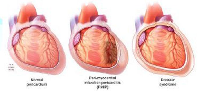
MEDizzy
MEDizzy
DR
Dr Raja Nandhini over 1 year ago
Dressler syndrome

Dressler syndrome

Dressler syndrome is a secondary form of pericarditis that occurs in the setting of injury to the heart or the pericardium (the outer lining of the heart). It consists of fever, pleuritic pain, pericarditis and/or a pericardial effusion. Also known as postmyocardial infarction syndrome and the term is sometimes used to refer to post-pericardiotomy pericarditis. It was first characterized by William Dressler at Maimonides Medical Center in 1956. Usually evidenced by a pericardial friction rub, chest pain worsening when recumbent, and diffuse ST elevation with PR segment depression and/or a pericardial effusion. Very rarely leads to pericardial tamponade.Elevated ESR is an objective but nonspecific laboratory finding. Treatment will be high dose of aspirin, corticosteroids and NSAIDS

15
Other commentsSign in to post comments. You don't have an account? Sign up now!
Related posts
Fibrinopurulent pericarditis.Heart and the pericardiumHeart Valves and Cusps IIHeart Valves and CuspsHeart Attack Vs Cardiac ArrestOral Diuresis in Heart FailureMalabsorption syndromePeutz jeghers syndromeHeart and Lung Auscultation### Anatomy of the Heart: Summary The heart is a muscular organ responsible for pumping blood throughout the body, supplying oxygen and nutrients while removing waste products. It is located in the thoracic cavity, between the lungs, and is roughly the size of a fist. #### 1. **Structure:** - **Chambers:** The heart has four chambers: - **Right Atrium:** Receives deoxygenated blood from the body via the superior and inferior vena cavae. - **Right Ventricle:** Pumps deoxygenated blood to the lungs through the pulmonary arteries. - **Left Atrium:** Receives oxygenated blood from the lungs via the pulmonary veins. - **Left Ventricle:** Pumps oxygenated blood to the rest of the body through the aorta. - **Valves:** The heart contains four main valves that ensure unidirectional blood flow: - **Tricuspid Valve:** Located between the right atrium and right ventricle. - **Pulmonary Valve:** Located between the right ventricle and pulmonary arteries. - **Mitral (Bicuspid) Valve:** Located between the left atrium and left ventricle. - **Aortic Valve:** Located between the left ventricle and the aorta. #### 2. **Wall Layers:** - **Epicardium:** The outer layer, also known as the visceral pericardium. - **Myocardium:** The thick, muscular middle layer responsible for contraction. - **Endocardium:** The inner layer that lines the chambers and valves. #### 3. **Blood Supply:** - The heart receives its blood supply from the coronary arteries, which branch off the aorta. The **right coronary artery** supplies the right side of the heart, while the **left coronary artery** branches into the left anterior descending and circumflex arteries, supplying the left side. #### 4. **Electrical System:** - The heart's rhythmic contractions are controlled by its electrical conduction system, which includes: - **Sinoatrial (SA) Node:** The natural pacemaker located in the right atrium. - **Atrioventricular (AV) Node:** Receives impulses from the SA node and transmits them to the ventricles. - **Bundle of His and Purkinje Fibers:** Distribute the electrical impulse throughout the ventricles, leading to contraction. #### 5. **Function:** - The heart functions in two primary circulatory loops: - **Pulmonary Circulation:** Moves deoxygenated blood from the right side of the heart to the lungs for oxygenation. - **Systemic Circulation:** Distributes oxygenated blood from the left side of the heart to the rest of the body. The anatomy of the heart is crucial for its role in maintaining effective circulation and overall cardiovascular health. Understanding its structure helps in diagnosing and treating various heart conditions.

Recent MCQs


Show more MCQs

Recent flashcard sets


Show more flashcards