Endocardial Calcification in Behçet's Disease
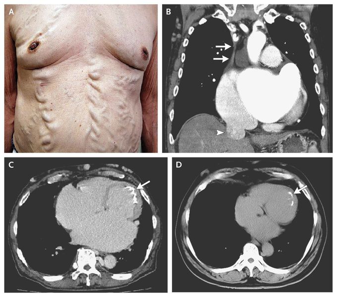
A 68-year-old man who had Behçet's disease with a 30-year history of oral and genital ulcers and erythema nodosum presented with progressive leg edema and dyspnea. The physical examination revealed varices of the chest and abdominal wall (Panel A), caused by collateral circulation associated with obstruction of the superior vena cava (SVC). Chest computed tomography (CT) showed SVC obstruction (Panel B, arrows) and dilatation of the inferior vena cava (arrowhead). Endocardial calcification on the midlateral and apical wall (Panel C, arrow) was more extensive than that seen on imaging performed 11 years earlier (Panel D, arrow), and there was increased dilatation of the right ventricle and both atria. A restrictive cardiomyopathy with endomyocardial fibrosis, which can be a complication of Behçet's disease, was diagnosed on the basis of echocardiography (video) and cardiac catheterization (including endomyocardial biopsy). After diuresis, the edema and dyspnea improved, but the varices of the trunk and lower limbs remained. Takafumi Nishida, M.D. Daihiko Hakuno, M.D., Ph.D. National Defense Medical College, Saitama, Japan source: nejm.org


Thanks for the case. I've been studying vasculitis these days, and it was really enriching for me.