


A Sublingual Epidermoid Cyst
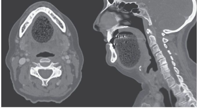
A 73-year-old woman presented to the emergency department with difficulty in speaking, which had progressively worsened over a period of 4 days. Her medical history was notable only for a dental procedure 2 months earlier that had involved the insertion of a bridge because of a missing tooth. Physical examination showed a large, fluctuant, painless swelling of the anterior floor of the mouth. There were no palpable cervical lymph nodes and no purulent drainage at the orifices of the submandibular and sublingual ducts. Computed tomography of the neck with the use of contrast material revealed a well-defined, midline, and suprahyoid cyst, measuring 7 cm by 4 cm by 3.5 cm, located above the geniohyoid and mylohyoid muscles (the image on the left shows the axial view, and the image on the right the sagittal view). The lesion had peripheral enhancement with multiple discrete foci of hypoattenuation indicating a coalescence of small, fatty nodules — features consistent with an epidermoid cyst. Epidermoid cysts are benign cysts filled with keratin. Although most commonly located on the face, neck, and trunk, they can be present within the floor of the mouth, as in this case. This patient’s cyst was surgically excised with an intraoral approach, and she had complete resolution of her symptoms.
could someone describe/explain how would purulent drainage look like?
