


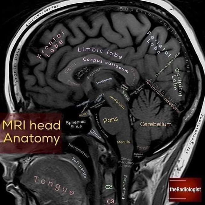
MRI Head Anatomy
Being a Doctor or future Doctor, always remember that our basics should be clear enough to diagnose and treat our patients. Here I'm sharing u some series of X-rays/ MRI/ CT scan with the location of different parts of our internal body system.

Report Date CPT: MRI BRAIN WITHOUT CONTRAST HNIQUE: MRI brain with contrast REPORT: large altered signal intensity area measuring 8.7x6.6cm having solid and cystic component is seen in right frontal lobe appearing hypointense on T1 and heterogeneously hyperintense on T2 and FLAIR images. The solid component reveals patchy diffusion restriction on DWI and patchy heterogeneous contrast enhancement on post contrast images. MRS reveals markedly elevated choline peak with reversed choline/creatine ratio and markedly reduced NAA. There is subtle surrounding edema especially posteriorly. The lesion is having mass effect on the ipsilateral ventricle with midline shift. Extension of the lesion to the corpus callosum and across the midline is also noted. Rest of the brain parenchyma is unremarkable. SION: Findings are suggestive of large butterfly glioma in right frontal lobe. RUKHSANA AZIZ
