


Adamantinomatous Craniopharyngioma Containing Teeth
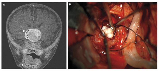
A 4-month-old term male infant presented with increasing head circumference on routine pediatric visits. Magnetic resonance imaging (MRI) of the brain revealed a heterogeneous, enhancing suprasellar mass (4.1 cm by 4.0 cm by 3.5 cm) on T1-weighted images after the administration of contrast material (Panel A). Multiple structures along the right periphery of the mass showed characteristics similar to those of teeth in the mandible (arrows). The patient underwent tumor resection through a right pterional approach; multiple fully formed teeth were seen in the tumor mass (Panel B). On pathological examination, an adamantinomatous craniopharyngioma was identified, which is a slow-growing tumor arising from Rathke's pouch, an embryonic precursor to the anterior pituitary. Such tumors consist of organized stratified squamous epithelium and wet keratin (keratin nodules) and may be cystic; the cysts are filled with viscous, yellow fluid containing cholesterol crystals. Histologically, adamantinomatous craniopharyngiomas closely resemble some odontogenic tumors and cysts. In the year since he underwent surgery, the patient has required shunting for bilateral subdural hygromas and receives thyroid and adrenal hormone-replacement therapy. He is making good developmental progress, and as part of his follow-up, he currently undergoes routine MRI.
