


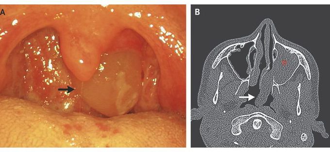
Intraoral Presentation of Antrochoanal Polyp
An otherwise healthy 17-year-old boy presented for evaluation of an incidentally noticed pharyngeal mass. He did not have nasal obstruction, rhinorrhea, epistaxis, or postnasal drip. Intraoral examination showed a soft, translucent, round mass occupying the left pharyngeal area (Panel A, arrow). Rhinoscopy and computed tomography of the sinuses revealed an antrochoanal polyp originating from the left maxillary sinus (Panel B, asterisk), extending through the accessory ostium into the middle meatus and then out posteriorly through the choana into the oropharynx (Panel B, arrow). The polyp was completely removed by means of endoscopic maxillary sinus surgery while the patient was under general anesthesia. The histologic diagnosis was consistent with a benign inflammatory polyp. At follow-up 6 months after surgery, there were no signs of recurrence.
