

Chronic Obstructive Pyelonephritis
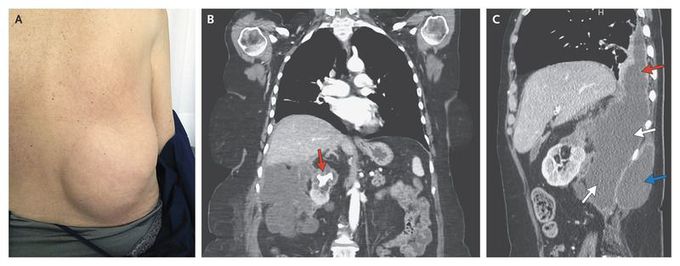
A 65-year-old woman presented to the hospital with a 4-month history of intermittent low-grade fever, asthenia, anorexia, and weight loss. Her medical history was limited to atrophic gastritis treated with intramuscular vitamin B12. Physical examination revealed a large, soft mass protruding from her right flank (Panel A, view from posterior). Blood tests showed a leukocyte count of 17,900 per cubic millimeter (90% neutrophils), a hemoglobin level of 9.8 g per deciliter, a C-reactive protein level of 274 mg per liter, and an erythrocyte sedimentation rate of 104 mm per hour. Blood cultures were negative. Computed tomography of the abdomen with the use of contrast material revealed a large, obstructing calculus in the upper-pole calyx of the right kidney, causing focal atrophy and cortical thinning of the upper pole (Panel B, arrow); there was also a large retroperitoneal abscess (Panel C, white arrows) extending into the chest (Panel C, red arrow) and subcutaneous tissues of the right flank (Panel C, blue arrow). A urine culture was positive for Proteus mirabilis, as was a culture of fluid drained percutaneously from the abscess. A right nephrectomy was performed. Histologic examination revealed an abscess with chronic inflammation and no xanthogranulomatous changes. The patient was doing well 18 months after surgery.

