


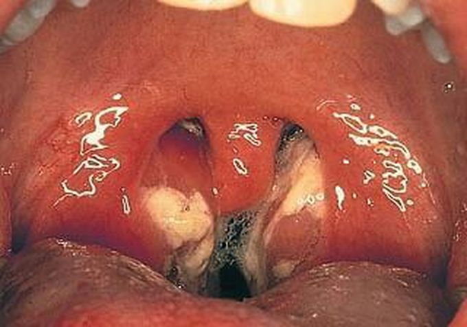
Infectious mononucleosis ( kissing disease) from its transmission by saliva) is an infectious, widespread viral disease most commonly caused by the Epstein–Barr virus (#EBV) It causes symptoms simillar to staphylococcus infection and leukemia Do you know how we can tell the difference between mononucleosis and leukemia? #medical #medstudent #premed #premedlife #nurse #kissing #disease #doctor #nurse #mononucleosis #throat #blood #saliva #pathology #disease #hospital #downey #ebv #virus #infection
 Mono vs. acute leukemia May 5, 2011 | acute leukemia, benign leukocytoses, Hematopathology  Q. I’m a medical student, and I wonder if you have some good tips about how to tell tell apart mononucleosis from AML on a blood smear? A. Great question! It would obviously be important to be able to distinguish between infectious mononucleosis (a benign disorder caused by EBV) and acute myeloid leukemia (a malignant disorder characterized by proliferation of myeloblasts). I have some general guidelines that I would use to tell those two entities apart, but first let’s review what you’d see in each disorder. Infectious mononucleosis is characterized by an elevated white blood cell count. The majority of the white cells are lymphocytes which have an unusual appearance. These are called “reactive lymphocytes” and they appear quite different from normal lymphocytes. There are three types of reactive lymphocytes commonly seen in mono: the Downey I lymphocyte (a small lymphocyte with lobed nucleus and scant cytoplasm), the Downey II cell (a large lymphocyte with copious cytoplasm that reaches out and abuts adjacent cells), and the Downey III cell (a large lymphocyte with finely reticular chromatin and abundant cytoplasm – there’s one in the photo above). Acute myeloid leukemia is also characterized by an elevated white blood cell count. The majority of the white cells are usually myeloblasts (the earliest precursors in the myeloid cell series), which are large cells with a high nuclear/cytoplasmic ratio and very fine chromatin. Usually you’ll be able to see some nucleoli, and sometimes you can find unique structures called Auer rods (which only exist in malignant myeloid cells). If you have a blood smear with an elevated white blood cell count, and you see lots of big cells with fine chromatin, it could be difficult to tell the two apart! Here are some thoughts as to how you might distinguish the two: 1. First, take a look at the total white count. In infectious mononucleosis, the white count is usually moderately elevated, usually somewhere in the range of 16-20 (normal for adults is around 5-11). It rarely goes above 35. In AML, however, the WBC is usually markedly elevated (50, or even 100 or more). Granted, if you catch the AML early in its course, the white count may be lower, but that’s a rare occurrence. So the magnitude of the leukocytosis is usually helpful. 2. Second, take a look at the cells themselves. In infectious mono, the lymphocytes are generally pretty heterogeneous. There will usually be a lot of Downey II cells, with smaller numbers of Downey I and Downey III cells. In AML, most of the white cells will be blasts (at least 20% of the nucleated cells in the blood or bone marrow must be blasts in order to call
