


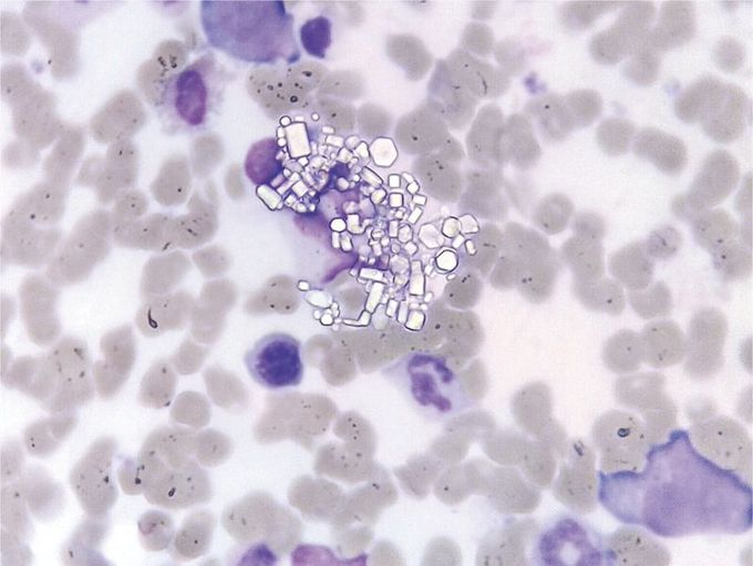
Cystine Crystals in Bone Marrow
A 1-year-old girl presented with a history of growth failure, mild anemia, hypophosphatemia, hypovitaminosis D, glucosuria, and proteinuria. She underwent diagnostic bone marrow aspiration, which revealed an increased proportion of macrophages containing polygonal crystals (20%; normal range, 2 to 5%), which are pathognomonic for cystinosis. A rare autosomal recessive disorder, cystinosis is caused by a defect in lysosomal storage of the amino acid cysteine, typically owing to a mutation in cystinosin, a lysosomal membrane protein. Free cystine accumulates in lysosomes in cells, including cells in the kidneys, liver, eyes, and brain. The diagnosis is made by observing cystine crystals or by measuring the cystine content of leukocytes. Although cystine crystals that are observed in the cornea on slit-lamp examination or in the bone marrow are diagnostic of cystinosis, they may be absent in patients younger than 1 year of age. The child began cysteamine treatment and had improvement in clinical symptoms; however, 8 months after the diagnosis of cystinosis, the child died from a generalized infection during a hospital admission.
