


Knee Dislocation - Everything You Need To Know - Dr. Nabil Ebraheim
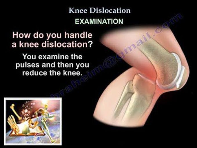
Dr. Ebraheim’s educational animated video describes dislocation of the knee and types of knee dislocations. Dislocation of the knee is a serious problem. It should be recognized and managed appropriately early. Knee dislocation is considered an orthopedic emergency. It is important to recognize the dislocation, and do reduction and perform serial neurovascular exam before the reduction and after the reduction. Approximately 50% of knee dislocations spontaneously reduce before formal evaluation. Morbid Obesity can be a risk factor for knee dislocation. Because the joint capsule is torn, knee swelling may not occur with knee dislocation. The knee dislocation associated with sports injuries have a lower incidence of neurovascular injury than those associated with a high energy mechanism. There are several types of knee dislocation that are usually described in the literature. There is a classification based on direction of displacement of the tibia and classification system based on the severity of the ligamentous damage was developed by Dr. Schenck. The dislocation of the knee usually involves at least two ligaments. Anterior knee dislocation is caused by hyperextension mechanism that causes failure of the posterior capsule, the PCL and sometimes the ACL. Anterior knee dislocations are the most common types. Posterior dislocations are seen in dashboard injuries. The posterior dislocation has the highest rate of vascular injury (about 25%), and sometimes will be a complete tear of the popliteal artery. Lateral dislocation has the highest rate of peroneal nerve injury. Posterolateral dislocation is the most common rotatory dislocation and it is usually irreducible. In rotatory dislocations, the PCL remains intact as the tibia rotates about the femur. A dimple on the medial side indicates posterolateral dislocation, which means that the dislocation cam be irreducible and the medial femoral condyle button hoes through the joint capsule. The physical examination and diagnostic studies can direct the treatment in a timely fashion. There could be an obvious deformity. See if there is a dimple sign. You need to check the pulses, get the x-ray, and reduce the knee immediately regardless if there is a pulse or no pulse. When you check the pulses, compare it to the other side. You have to do serial exam of the pulses and the whole idea is to make sure to discover if there is a vascular injury or not, because if there is a vascular injury and there is a delay more than 8 hours in reestablishing the arterial blood flow, that will result in an amputation rate of 85%. The patient may have severe pain, instability, and the exam will be difficult because of guarding and apprehension, but keep focusing on the circulation. Look at the pulses. Look at the x-rays! The patient may have a fracture, such as medical tibial plateau fracture and other intraarticular fractures, the patient may have asymmetry of the joint space, avulsion fractures such as the Segond sign for ACL tear or tibial eminence avulsion fracture. Look for asymmetry of an irregular joint space on the x-rays. After you reduce, you immobilize, get x-rays, and make sure the dislocation is reduced. Up to 1/3 of knee dislocation have associated injuries that require more urgent attention. Knee dislocation is a high energy injury that will have popliteal artery injury, nerve injury, multiple fractures, head and chest trauma, and compartment syndrome. The findings of the knee might be subtle, but having knee dislocation makes you think of these conditions and the swelling may not be that significant because the joint capsule is torn. You may have hyperextension of the knee, popliteal ecchymosis, foot drop, or vascular issues, and the patient may also be normal. The knee can be dislocated or be normal. The patient may or not be overweight. You examine the pulses and then you reduce the knee. After you reduce the knee, you examine the pulses and get post reduction A.B.I. (ankle brachial index). The ankle brachial index (A.B.I.) is very important in the knee dislocation. The systolic blood pressure measured is divided by that measured at the brachial artery. A ratio less than 0.9 is considered abnormal and needs further investigation. Scenario 1: Good pulses and symmetrical, A.B.I. is more than 0.9, then observe for 24-48 hours with serial neurovascular exam. Scenario 2: After the reduction, if the distal pulses are asymmetrical, or the A.B.I. is less than 0.9, then you will do some kind of a study to see why this is occurring (get a CTA or arteriogram). Scenario 3: If you have absent distal pulses or clear, hard signs of limb ischemia, do not waste time by doing arteriogram. Take the patient to the operating room, do emergency exploration and then on the table, do arteriogram and the patient will probably need fasciotomy. You will stabilize the knee with and external fixator.
