


Froin’s Syndrome
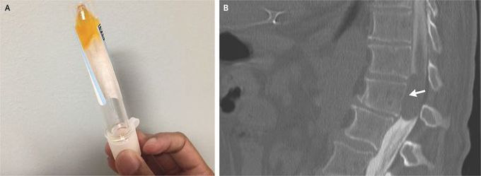
A 64-year-old man presented with a 1-week history of progressive bilateral weakness in the legs. The neurologic examination showed paralysis of the legs and decreased sensation starting at the L1–L2 level. Magnetic resonance imaging could not be performed owing to the presence of a pacemaker. Since the patient had atrial fibrillation and an elevated prothrombin time, there was concern about a possible spinal hematoma. Computed tomography (CT) of the spine showed only degenerative disk disease. On lumbar puncture, the cerebrospinal fluid (CSF) was xanthochromic, viscous, and coagulated in the tube (Panel A). The protein level in the CSF was more than 1500 mg per deciliter, and the glucose level was 45 mg per deciliter (2.5 mmol per liter). The CSF contained less than one nucleated cell per cubic millimeter, and the results of Gram’s staining and cultures were negative. The combination of elevated protein, xanthochromia, and hypercoagulation of CSF is pathognomonic for Froin’s syndrome, which can occur with blockage of CSF flow by a spinal cord mass or with meningeal irritation from meningitis. CT myelography showed a large intradural, extramedullary lesion at T11–T12 (Panel B, arrow), which was compressing the spinal cord. The patient underwent total laminectomy of T11 and T12 and partial laminectomy of L1 with tumor resection; a benign nerve-sheath tumor (schwannoma) was diagnosed on pathological analysis. No radiotherapy or chemotherapy was performed. After 1 month of rehabilitation, the patient had improved sensation but continued having leg paralysis.
