


Gartner’s Duct Cyst
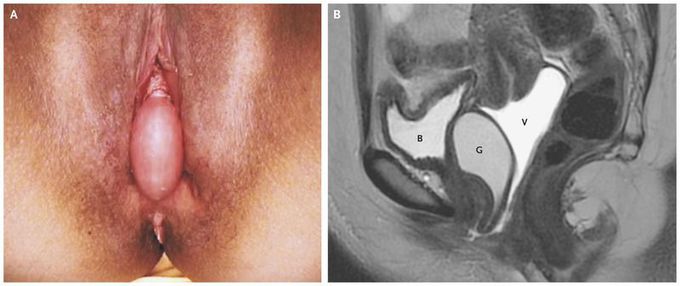
A 29-year-old woman presented to an outpatient gynecology clinic 4 months after an uncomplicated vaginal delivery with a mass protruding from the vaginal canal. She reported no urinary tract symptoms or abnormal vaginal discharge. A physical examination revealed a smooth, nontender mass, 5 cm in the longest diameter, in the midline of the anterior wall of the vagina (Panel A). There was no evidence of pelvic-organ prolapse. A cystoscopic examination did not reveal a urethral or bladder diverticulum. Magnetic resonance imaging of the pelvis (Panel B) with contrast medium inserted into the vagina revealed a cystic structure (G) located between the bladder (B) and the vagina (V). The patient underwent complete resection of the cyst, which was histologically confirmed to be a Gartner’s duct cyst. A Gartner’s duct cyst is a benign vaginal cystic structure that arises from the vestigial remnant of the mesonephric (wolffian) duct, which, during male embryogenesis, forms the seminal vesicles, vas deferens, and epididymis. Gartner’s ducts are paired structures on either side of the urethra; when cysts become large, as in this patient, they can appear in the midline. The patient had an unremarkable postoperative course and had no sign of recurrence 6 weeks after surgery.
