Intravenous Administration of Fluorescein in Brain-Tumor Surgery
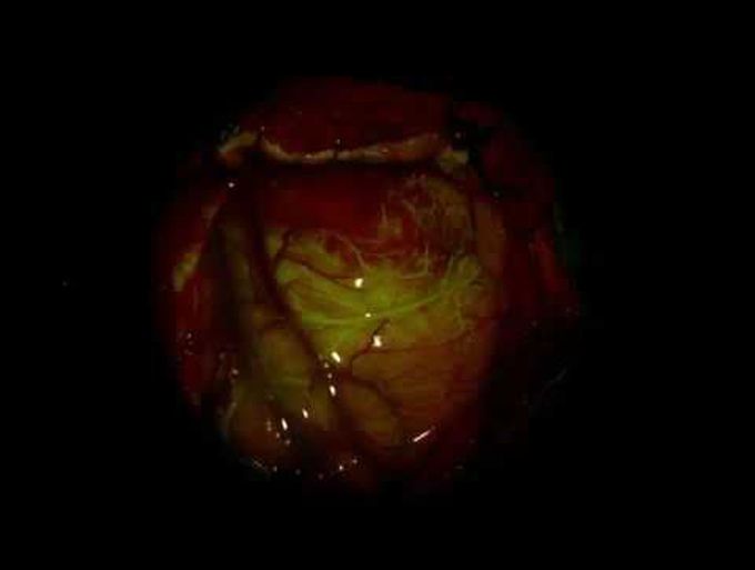
Fluorescein Guidance in Glioblastoma Resection A 55-year-old man presented to the emergency department with a 1-week history of severe headache. Magnetic resonance imaging of the brain with and without gadolinium-based contrast agent revealed a rim-enhancing mass (4.5 cm in diameter) in the right temporal lobe. At the time of surgery for tumor resection, fluorescein was administered intravenously (Video); Panel A shows the baseline condition before injection. The intact blood–brain barrier of normal brain parenchyma prevents the uptake of fluorescein; however, a tumor can disrupt the blood–brain barrier and allow for the accumulation of the fluorophore. In this patient, within the first 35 seconds after the injection, fluorescence was first evident in arterial vessels (Panel B), then in the smaller veins, and then in the larger veins (Panel C). By 35 seconds after the injection, fluorescein had begun to accumulate in the tumor tissue (Panel C). At 86 seconds, the fluorescein within the cortical veins had begun to fade, providing a sharp contrast between the tumor (Panel D, area inside the dashed line) and normal surrounding tissue; a small area of blood obscures some tumor tissue. Pathological studies revealed a glioblastoma, and postoperative magnetic resonance imaging confirmed gross total resection of the tumor. Four weeks after surgery, the patient began to receive external-beam radiation therapy and temozolomide chemotherapy. At follow-up 2 months after surgery, he had no symptoms of headache and did not have any neurologic deficits.