

Sturge–Weber Syndrome
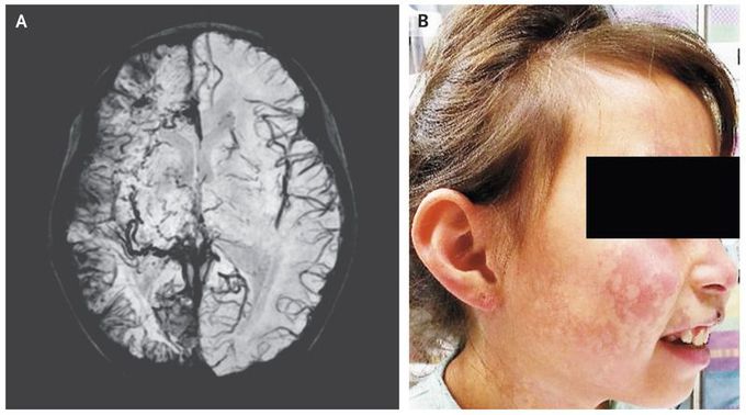
A 6-year-old girl was brought to the emergency department after the sudden onset of weakness in the left arm and leg. She also reported a 2-day history of pain in her right eye. No seizure activity had been witnessed. She had a nonpalpable red lesion on the right side of her face, and the intraocular pressure was elevated in both eyes. Magnetic resonance imaging (MRI) of the head revealed right cerebral atrophy with leptomeningeal enhancement along the right cerebral convexity. Susceptibility-weighted MRI (Panel A) revealed abnormal leptomeningeal capillary vessels along the right cerebral convexity. The MRI and clinical findings were consistent with the Sturge–Weber syndrome, a rare congenital vascular disorder characterized by a cutaneous capillary malformation, also called port-wine birthmark (Panel B shows the patient’s face 2 years after the initial presentation), and abnormal capillary venous vessels in the brain and eye that can lead to glaucoma, seizures, stroke, and intellectual disability. The weakness on the left side, which was thought to be postictal paresis, resolved within 24 hours. An antiepileptic medication was prescribed, as well as timolol and dorzolamide for glaucoma and daily low-dose aspirin treatment to reduce the risk of thrombotic events. Over the next 2 years, she had two presentations with seizures, which led to adjustment of the antiepileptic regimen.

