
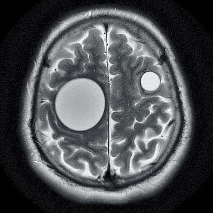
Hydatid cysts
This unique image shows perfectly round hydatid cysts within the brain of a 35-year-old male previously treated for a liver infection. The common cause is a parasitic infection by Echinococcus granulosus. The infection is acquired via contaminated food with eggs of the tapeworm. The oncospheres released from the eggs in the bowel enters the portal circulation. Hence the liver is most commonly affected, followed by the lung and other organs such as bones, genitourinary system, bowel and subcutaneous tissues.Hydatid cysts end up in the brain due to direct invasion of larva that managed and filtered via liver and lung to the brain. The MRI shows well-defined circumscribed spherical non-enhancing intra-axial cystic lesion that usually lies in the territory of the middle cerebral artery.Management is surgical, with removal of the entire cyst without rupture using Dowling’s maneuver (instilling warm saline between the cyst wall and the brain).Credit to @radiopaedia