


Medicaltalks almost 8 years ago
#NSFWSensitive content 18+
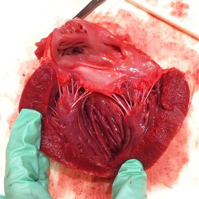
This material contains content which some users might find distrubingLeft ventricle after dissection
The following image shows the normal appearance of the left ventricle after dissection, sent by one of our followers (@danikalexis). The beautifully arranged tendinous chords (heart strings) are connecting the papillary muscles to the mitral valve (left ventricle) or to the tricuspid valve (right ventricle). That way they prevent backflow of blood into the atria during systole. The chords also prevent valve prolapse by becoming tense thus pulling the flaps, holding them in closed position.
Other commentsSign in to post comments. You don't have an account? Sign up now!
