


Melanoma Metastatic to the Duodenum
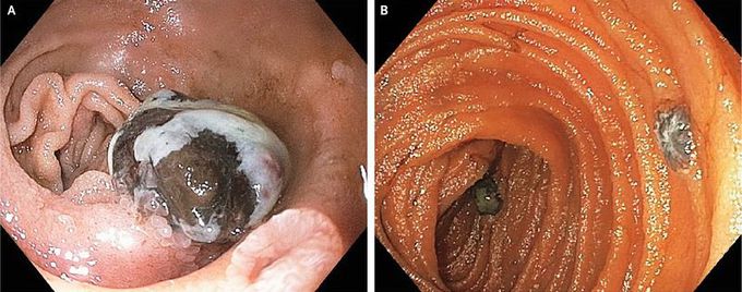
An 87-year-old man presented to the emergency department with a 3-week history of progressive nausea, intermittent coffee-ground emesis, anorexia, and weight loss. The patient had a history of type 2 diabetes mellitus, peripheral artery disease, and aortic-valve replacement. Laboratory evaluation showed microcytic anemia and severe iron deficiency. Upper endoscopy revealed a pigmented mass in the duodenal bulb (Panel A) and two additional masses in the second and third portions of the duodenum (Panel B). Biopsies of these lesions confirmed a diagnosis of melanoma. The patient had received no prior diagnosis of cutaneous melanoma. Positron-emission tomography–computed tomography revealed fluorodeoxyglucose uptake in the duodenum, omentum, and subcarinal, retroperitoneal, and superficial inguinal lymph nodes, findings that are consistent with widely metastatic disease. Melanoma can metastasize to many organ systems, including the gastrointestinal tract. After the diagnosis was made, the patient’s care was transferred to an oncology center for further treatment.
