


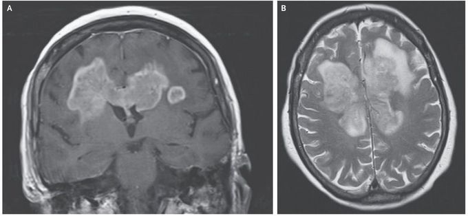
Butterfly Glioma
An 83-year-old woman presented to the emergency department with a 5-hour history of weakness in the left arm, drooping on the right side of the face, slurred speech, and urinary incontinence. A 7-week history of progressive functional and cognitive decline was reported. Gadolinium-enhanced magnetic resonance imaging (MRI) of the head showed a solid, enhancing lesion on the T1-weighted sequence, with a butterfly appearance that extended across the corpus callosum (Panel A). Hyperintensity of the mass was observed on the T2-weighted sequence (Panel B), and restricted diffusion was seen on diffusion-weighted MRI. The differential diagnosis for lesions that extend across the corpus callosum and symmetrically into the frontal lobes includes glioblastoma and primary central nervous system lymphoma. A biopsy of the right frontal lobe revealed a glioblastoma. After discussion regarding the limited treatment options, including radiation and chemotherapy, for this extensive tumor, the patient and family opted for supportive, palliative management. The patient died 3 weeks later.
