

Coronary Arteritis in IgG4-Related Disease
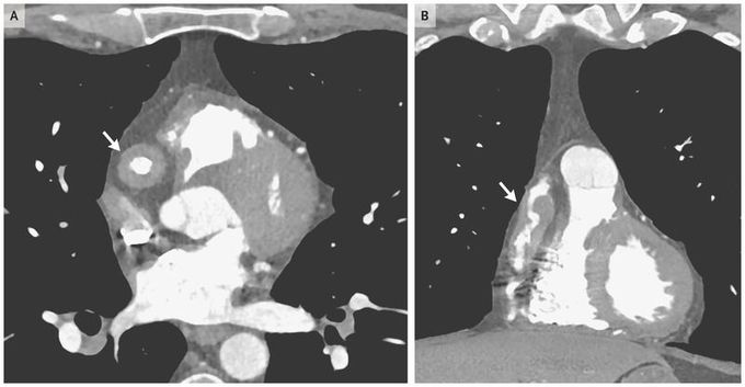
A 47-year-old man presented to the cardiology clinic for evaluation of coronary-artery dilatations that had been detected on computed tomography (CT) of the chest performed as a follow-up examination of pulmonary nodules. The patient was asymptomatic but had a history of biopsy-proven IgG4-related disease involving the pancreas and the lacrimal and parotid glands. At the current presentation, he had no swelling of the lacrimal or parotid gland or abdominal tenderness, all of which had characterized previous disease flares. He had been receiving rituximab for the past 4 years. Results of laboratory tests showed a normal erythrocyte sedimentation rate and serum IgG4 concentration. An electrocardiogram-gated CT angiogram of the coronary vessels revealed aneurysmal dilatation of the right coronary artery with marked, nearly circumferential, periarterial soft-tissue thickening in the transverse view (Panel A, arrow) and the coronal view (Panel B, arrow). Diffuse thickening and aneurysmal changes were also seen in the left anterior descending coronary artery. The thoracic aorta, branch vessels of the aortic arch, and abdominal aorta showed no evidence of disease. It was assumed that these abnormalities occurred during the earlier period of active disease and represented damage rather than active IgG4-related disease. Treatment with low-dose aspirin was started. At a follow-up examination 4 months later, the patient remained asymptomatic.

