


Pulmonary Metastases from Chondroblastic Osteosarcoma
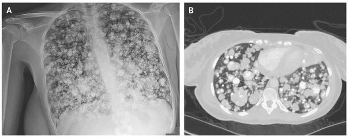
A 21-year-old woman presented to the emergency department with an 8-month history of progressive swelling and pain in the right thigh. Magnetic resonance imaging of the right leg revealed a large, enhancing, circumferential periosteal femoral mass that measured 30 cm in the craniocaudal dimension, as well as a nonocclusive thrombus in the right femoral vein, for which the patient received anticoagulation. Biopsy of the femoral mass confirmed a diagnosis of chondroblastic osteosarcoma. One week later, the patient had shortness of breath and left pleuritic chest pain. The oxygen saturation was 94% while the patient was breathing ambient air. Chest radiography revealed innumerable lesions in both lungs (Panel A). Computed tomography of the chest confirmed the presence of extensive, lobulated, and partially calcified nodules and masses throughout both lungs, a finding suggestive of pulmonary metastases (Panel B). Despite the initiation of chemotherapy, the disease progressed. A second chemotherapy regimen was started; five cycles have been completed, and the disease is currently stable.
