


Vertebral Hydatidosis
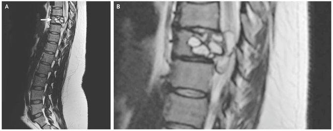
A 35-year-old woman presented to the emergency department with weakness, a feeling of electric shocks in both legs, and repeated falls. She reported that the symptoms had been progressing, and she noted that she had had difficulty riding her horse for the preceding 3 months. She lived in France, had no history of foreign travel, owned a pet cat, and had contact with cattle. Physical examination revealed impaired sensation in both legs and weak foot flexion. Laboratory tests showed an elevated total white-cell count (18,800 per cubic millimeter; reference range, 4000 to 10,000) with a normal absolute eosinophil count and a C-reactive protein level of 49 mg per liter (reference value, <5). Magnetic resonance imaging of the spine revealed a lobulated lesion of the 9th thoracic vertebra with an epidural component (Panel A, arrow; a close-up image is shown in Panel B). Surgical treatment involved posterior corpectomy of the 9th thoracic vertebra, osteosynthesis of the 7th through 11th thoracic vertebrae, and removal of the epidural lesion. Pathological testing revealed cystic echinococcosis (hydatidosis), and reverse-transcriptase–polymerase-chain-reaction assay identified Echinococcus granulosus. Echinococcus is a parasitic cestode that can infect dogs and other pets and farm animals, with humans as incidental hosts. Infection can cause cystic lesions in the liver and lungs and also in the central nervous system and bones. In addition to surgery, this patient was treated with the antiparasitic medication albendazole. At follow-up 9 months after presentation, the patient had no residual symptoms or sign of recurrence.
