


Bitot’s Spots
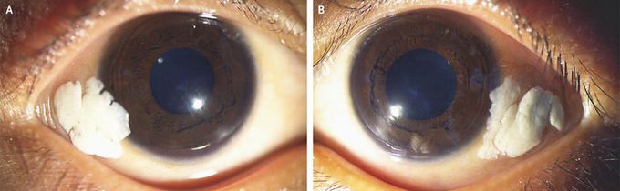
A 4-year-old boy was brought by his father to the ophthalmology clinic with a 1-year history of enlarging white deposits in both eyes and decreased night vision. On examination, the conjunctivae of both the right eye (Panel A) and the left eye (Panel B) appeared dry and wrinkled, with foamy, cream-colored deposits near the outer corners. The corneas were clear, the fundi were normal, and the visual acuity was 20/30 in both eyes. The child appeared pale, with hypopigmented hair, a weight of 10.5 kg (z score of less than −3), and a height of 92 cm (z score of −2.8). The ocular findings were consistent with Bitot’s spots, which are accumulations of keratin, often intermixed with an overgrowth of Corynebacterium xerosis, that result from epithelial metaplasia caused by vitamin A deficiency. Vitamin A deficiency can also cause blindness, as a result of corneal ulceration with scarring, and particularly night blindness, as a result of dysfunction of rod photoreceptor cells. In this patient, the serum vitamin A level was 16.8 μg per deciliter (0.59 μmol per liter) (reference range, 20 to 40 μg per deciliter [0.70 to 1.40 μmol per liter]), and the retinol-binding protein level was 0.01 g per liter (reference range, 0.03 to 0.06). Further history taking revealed extreme poverty, and further examination showed signs of dietary deficiency, with no evidence of intestinal parasites. The patient received an oral dose of vitamin A, which was repeated 4 weeks later. Artificial tears were also administered. The Bitot’s spots abated, although they did not completely resolve, over the course of 12 weeks. The parents were advised to administer another dose of vitamin A every 6 months until the child was 5 years of age.
Pinguecula A pinguecula (plural pingueculae) is very similar to a pterygium, and the two are often confused. However, a pinguecula occurs only on the conjunctiva (the thin, protective membrane that covers the surface of the eye), and will not grow across the cornea. Pinguecula symptoms It has very similar symptoms to a pterygium. It usually appears as a creamy-coloured or chalky growth on the white of the eye, between the eyelids. A pinguecula will also normally occur in the corner of the eye, near the nose, and can affect one eye or both. Just like a pterygium, a pinguecula can cause irritation, as well as difficulty wearing contact lenses. However, a pinguecula cannot grow across the cornea, and therefore will not affect vision. In some cases though, a pinguecula can become a pterygium, involving the cornea. Causes of pingueculae Just like pterygia, pingueculae generally occur between the ages of 20 and 50. They are also thought to be caused by environmental factors, such as climate, dust and UV light. Treating a pterygium or pinguecula There a number of different treatments for a pterygium or pinguecula. Normally, pterygium surgery will only be undertaken if the pterygium has troublesome symptoms, or is affecting vision. Otherwise, management with eye drops is often effective. Pingueculae are rarely surgically removed, and are usually treated with eye drops. However, if the pinguecula turns into a pterygium, surgery may be the best course.
