

Atrial Myxoma
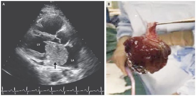
A previously healthy 47-year-old woman presented to her primary care physician with a 6-month history of worsening exertional dyspnea, progressive fatigue, and orthopnea. The physical examination was notable for sinus tachycardia, an abnormal heart sound initially thought to be an S3 gallop, and edema of both legs. A chest radiograph showed pulmonary venous congestion. Transthoracic echocardiography revealed a mass that was attached to the interatrial septum, with partial prolapse into the left ventricle obstructing the mitral-valve inflow during diastole (Panel A, arrow [LA denotes left atrium, and LV left ventricle],. The additional heart sound after the S2 was recognized as a characteristic “tumor plop.” The patient underwent excision of the left atrial mass (which measured 5.7 cm by 4.3 cm by 5.0 cm) (Panel B), resection of the interatrial septum, and reconstruction with a bovine pericardial patch. Pathological analysis confirmed the diagnosis of an atrial myxoma. Cardiac myxomas are the most common type of primary cardiac tumor in adults and usually occur in the left atrium. If the condition goes untreated, complications such as congestive heart failure, embolic stroke, and sudden death can occur. Surgical resection is the indicated therapy. A postoperative echocardiogram showed a normal ejection fraction and no flow across the interatrial septum. The patient had an unremarkable postoperative course and was discharged home.

