

Laryngocele
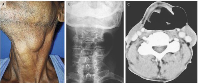
A 58-year-old man presented to the otorhinolaryngology outpatient clinic with a 2-year history of progressive hoarseness and swelling on the left side of his neck. He had no associated dysphagia, regurgitation of food, or dyspnea. He worked as a farmer and had no history of tobacco use. On physical examination, he had nontender, compressible swelling in the left cervical region that transmitted light on transillumination (Panel A). The swelling was accentuated when the Valsalva maneuver was performed Examination with a flexible fiberoptic laryngoscope showed a bulge over the left false vocal cord that was partially obstructing the airway lumen . A diagnosis of laryngocele was confirmed by radiography of the neck (Panel B) and by computed tomography (Panel C), both of which showed a well-defined lobulated structure located in the left paralaryngeal space and extending through the thyrohyoid membrane. A laryngocele is a dilatation of the laryngeal saccule within the sinus of Morgagni, the space between false and true vocal cords. It can result from activity that increases intralaryngeal pressure, such as excessive coughing, straining, playing a wind instrument, or glass blowing. The patient underwent a complete excision of the laryngocele. He remained asymptomatic at a follow-up visit 8 months later.

