


Cardiac Metastases in Melanoma
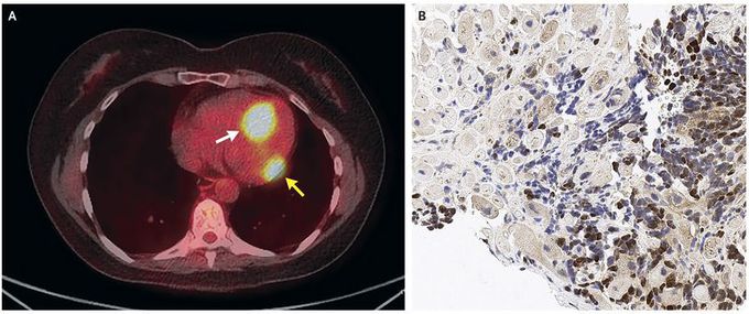
A 61-year-old woman was seen in the oncology clinic for evaluation of two cardiac masses found on annual surveillance with 18F-fluorodeoxyglucose–positron-emission tomography (FDG-PET) (Panel A). She was asymptomatic. Ten years earlier, biopsy of the nail bed of her left great toe revealed melanoma, which was surgically excised. She had local recurrence 7 years later, at which time she underwent amputation of the toe and received adjuvant treatment with ipilimumab. The current FDG-PET surveillance revealed FDG-avid masses within the left ventricle (Panel A, yellow arrow) and near the interventricular septum (Panel A, white arrow). No other FDG-avid sites were observed. Transthoracic echocardiography and computed tomography confirmed the presence of these two intramural lesions. An endovascular cardiac-biopsy specimen revealed melanoma metastatic to cardiac muscle, with tumor cells that were positive for a panel of melanocytic markers, including SOX10 (Panel B). Metastases to the heart from melanoma are rare, and cardiac involvement with no other sites of metastatic disease is rarer still. The patient started treatment with pembrolizumab, and follow-up imaging showed complete resolution of the cardiac metastases, with no new sites of disease. At follow-up 15 months after diagnosis, the patient continued to take pembrolizumab, with no recurrence of metastatic disease.
