


Visceral Leishmaniasis
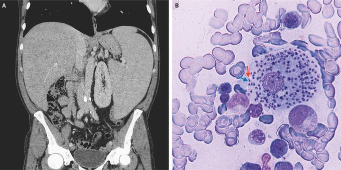
A 49-year-old previously healthy man presented to the emergency department with a 5-month history of fever, abdominal pain, fatigue, and an unintentional 15-kg weight loss. The physical examination was notable for an enlarged liver and spleen. Laboratory studies showed a white-cell count of 2040 per cubic millimeter (reference range, 4000 to 10,000), a hemoglobin level of 9.2 g per deciliter (reference range, 14.0 to 18.0), and a platelet count of 50,000 per cubic millimeter (reference range, 140,000 to 400,000). Blood cultures as well as tests for human immunodeficiency virus, hepatitis B virus, hepatitis C virus, cytomegalovirus, and Epstein–Barr virus were negative. Computed tomography of the abdomen confirmed the presence of an enlarged liver and markedly enlarged spleen (Panel A). Examination of a bone marrow aspirate revealed amastigotes, each with a nucleus (Panel B, blue arrow) and a kinetoplast (Panel B, red arrow), within histiocytes. This is the typical appearance of leishmaniasis, and polymerase-chain-reaction testing of the bone marrow aspirate confirmed the diagnosis. Transmitted by sandflies, Leishmania infantum is endemic to Italy and the Mediterranean region. The patient started treatment with liposomal amphotericin B. At a follow-up visit 1 month later, the fever, abdominal pain, and fatigue had resolved, and physical examination revealed resolution of the splenomegaly.
I was diagnosed as a Hepatitis B carrier in 2015, with early signs of liver fibrosis. At first, antiviral medications helped control the virus but over time, resistance developed, and the effectiveness faded. I began to lose hope. In 2021, I discovered NaturePath Herbal Clinic despite my skepticism, I decided to give their herbal treatment a try.To my surprise, after just six months, my blood tests came back negative for the virus.It was nothing short of life-changing.I never expected such incredible results from a natural treatment. But it not only cleared the virus it restored my hope, my health, and my peace of mind.If you or someone you know is battling Hepatitis B, I truly encourage you to explore the natural healing path offered by NaturePath Herbal Clinic. It gave me a second chance and it might do the same for you.www.naturepathherbalclinic.com info@naturepathherbalclinic.com
