


Andy Wellsabout 8 years ago
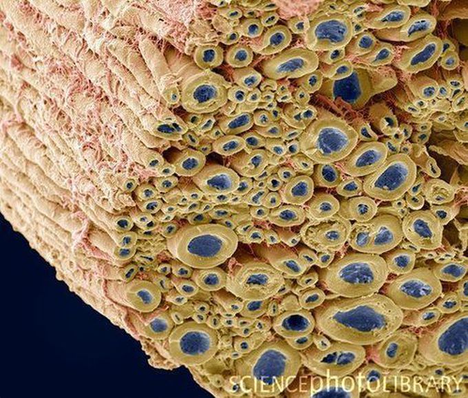
Nerve bundle
Nerve bundle. Coloured scanning electron micrograph (SEM) of a freeze-fractured section through a bundle of myelinated nerve fibres. Myelin sheaths (yellow) can be seen surrounding the axons (blue). Perineurium (connective tissue, pink) surrounds the nerve bundle while endoneurium divides the individual fibres.
Other commentsSign in to post comments. You don't have an account? Sign up now!
