

Laparoscopic management of urachal cysts
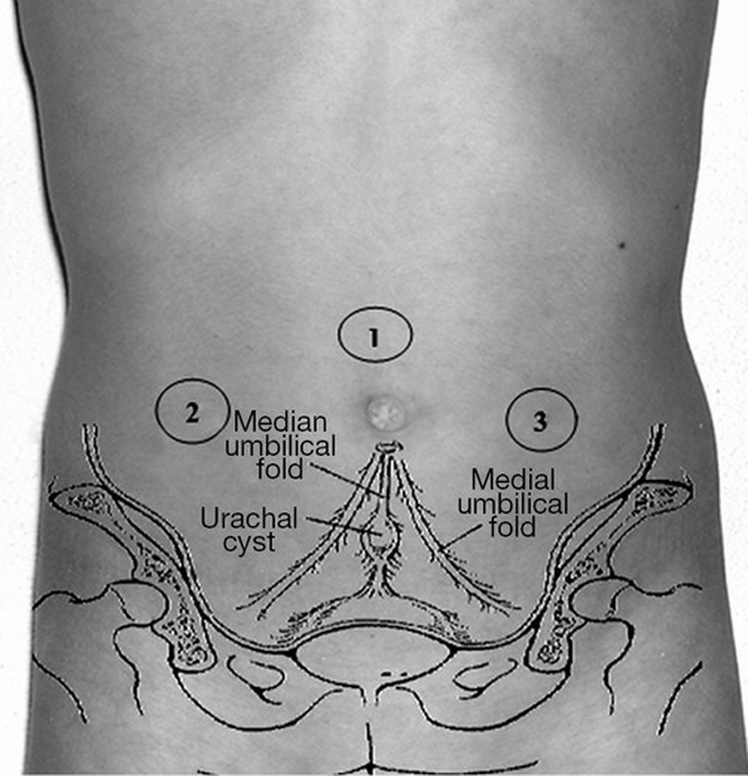
We included in this retrospective study all patients treated in our Department for urachal anomalies between 2006 and 2016 focusing our attention on urachal cysts diagnosis and mini-invasive treatment. This period was considered because in 2006 we started to treat this pathology laparoscopically. In a total of 16 patients 8 (50%), (6 females, 2 males) underwent open excision of an urachal remnant. The average age was 5.5 years (range, 4 months–13 years). The other eight patients were treated by laparoscopic surgery. We reviewed the charts of this group of patients to determine: age at surgery, preoperative imaging results, operative time, need for conversion, hospital stay length, postoperative complications. In this group signs and symptoms at onset were abdominal pain (acute in one patient and recurrent in two ones), macrohematuria and dysuria (1 pt), umbilical urinary discharge (3 pts), omphalitis with urinary umbilical discharge (1 pt). All patients underwent ultrasound, urinalysis was performed in 3 patients, while one of the eight was studied also with voiding cysto-urhethrogram and renal scintigraphy. The suspect of urachal remnant was based on the clinical symptoms and signs. All patients were operated on laparoscopically. Surgical technique: all patients were administered a single dose of parenteral Amoxicillin/Sulbactam during induction of anesthesia. The patient is placed in supine position with both arms secured at the side of the body. Foley’s catheterization of bladder was performed to initially decompress the bladder during the dissection of the urachal remnant and later allows a retrograde distension of the bladder at the time of wire the insertion of the urachus at the dome of the bladder. The operating table should be preferably inclined at 20°–30° in Trendelenburg position to allow the abdominal contents to fall in a cephalad manner away from the pelvis. The surgeon and the camera surgeon stand on the right side of the patient (at the patient’s head in younger ones) with the monitor in the opposite side of the patient (normally at the foot). Trocars position: the first 5 mm trocar can be placed with “open” technique infraumbilically and if this is not possible in the sovraumbilical region. Once the port is placed inside the peritoneal cavity, is created an appropriate pneumoperitoneum with CO2 pressures, which range from 6–8 to 12 mmHg according to the patient’s weight. A 5-mm 0° or 30° angled lens camera (according the surgeon’s preference) is inserted and under direct vision other two 3-mm/5-mm trocars are placed respectively in the right and left iliac fossa. A third accessory trocar can be placed in case

