


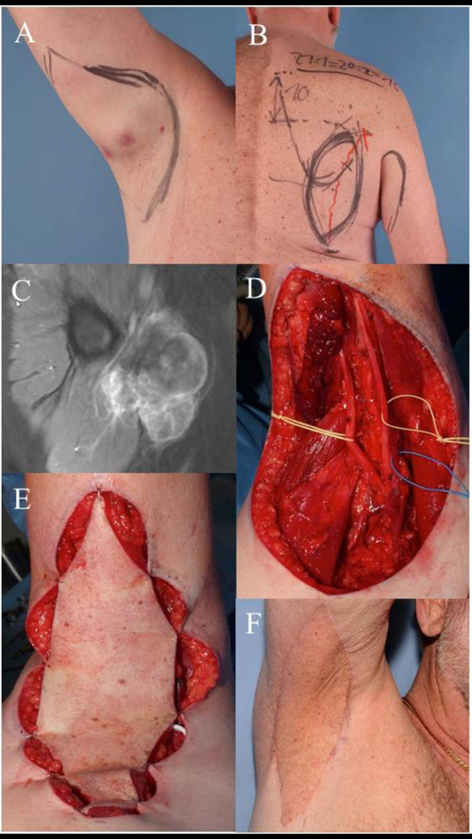
Case 1: (A) clinical visible mass in the right axilla, pre-OP drawing of the wide resection margins. (B)
Pre-OP drawing demonstrates planned pedicled parascapular fasciocutaneous flap. (C)Pre-OP MRI of the right axilla. (D)Intraoperative situation after wide en-bloc excision of tumor. Deep margins were achieved by epineurectomy and adventitiectomy. (E) Pedicled parascapular flap inset reconstructs the defect. (F) Clinical appearance 5 years after surgery and external post-OP radiation therapy. demonstrates the case of a 64-year-old male patient suffering from a high-grade pleomorphic sarcoma in the right axilla (T2b, N0, M0, G3). A wide excision was planned and carried out with sufficient removal of skin and subcutaneous tissue due to the extracompartimental localization of the sarcoma. Margins to the deep structures of the axilla could be achieved by thorough dissection of the nerve and vessel sheaths. The transfer of a pedicled parascapular flap was performed in the same operation. Noteworthy, we dissect the pedicle completely back through the medial axillary gap, ligating the osseous branches of the circumflex scapular artery. The flap can be advanced through this tunnel, allowing a completely tension-free placement in the axilla. The patient is shown 5 years after surgery and post-OP radiation therapy with an excellent functional result. The parascapular flap delivers sufficient pliable soft tissue coverage avoiding functional impairment of shoulder movement (53–55).
