


Arterial supply of the Stomach | Anatomy
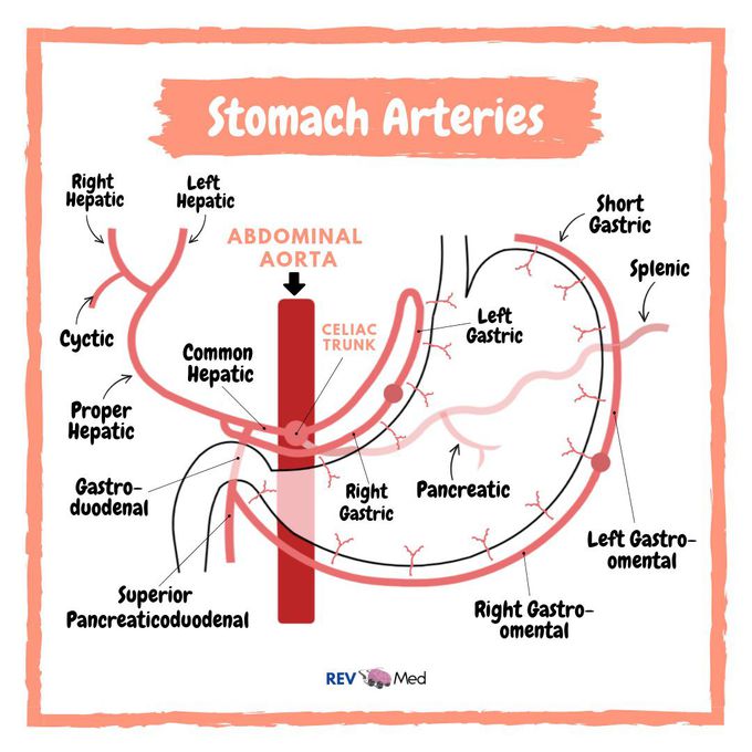
Link in bio for full video. Watch now! Notes below to follow this diagram, but I highly recommend you spend quick 4 mins and watch our video! The celiac trunk. It supplies the structures that are derived from the foregut: the stomach, proximal duodenum, spleen, liver, and most of the pancreas. Celiac Trunk (3 main arteries coming off of it) *Left Gastric - going up and looping *Common Hepatic - large going right *Splenic - tortuous going left to the spleen Look at the splenic artery first. The splenic artery follows a tortuous course towards the spleen along the upper border of the pancreas. Now let’s look to our right. The common hepatic artery passes and divides into the proper hepatic artery, and the gastro-duodenal artery. The Proper hepatic artery runs upwards and to the right, to supply the liver. Now look back at the common hepatic artery. From here this division two branches to the stomach arise, the right gastric artery, which usually arises from the hepatic artery, and the right gastro-omental (epiploic) artery, which arises from the gastro-duodenal. After giving off the right gastro-omental, the gastro-duodenal artery continues as the superior pancreatico-duodenal artery. It runs downward behind the duodenum, supplying it and the head of the pancreas. Now let’s take a look at the stomach, it’s divided into two arcades. First is right gastric (from hepatic artery) meeting with the left gastric (from celiac trunk), this occurs along the lesser curvature of the stomach. Second is the meeting of the right gastric-omental artery (from gastric-duodenal artery) with the left gastric-omental artery (from the splenic artery). Full Arterial Supply of Stomach video (link in bio) Instagram @rev.med
