


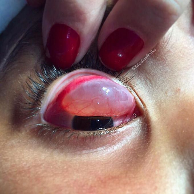
Giant conjunctival inclusion cyst bulging from underneath!
This is a benign cyst filled with clear serous fluid containing shed cells or gelatinous mucous material. Conjunctival inclusion cysts result from traumatic or iatrogenic implantation of conjuctival epithelial cells following trauma or surgery. However, the cysts can develop spontaneously or congenitally during the embryonic period by separation of a portion of conjunctival epithelial cells. The diagnosis of an inclusion cyst is usually made based on a typical translucent cystic appearance of the lesion. However, in cases where bleeding, infection or pigments are present in the intracystic materials, the definite diagnosis may be difficult by clinical examination alone. In this patient, the conjunctival cyst manifested atypically as a large clear, thin-walled mass extending from the conjunctival upper portion to the palpebral surface, and it’s pretty big. Remedy for cysts is complete excision. As the cysts are thin walled, rupture is common during excision. Recurrence is the main postoperative concern so careful and intact removal of cyst is necessary to prevent recurrence. Photo by @gustavo_da_paz

Hemodynamic stimuli&nonhemodynamic stimuliEffects of sugar on teeth
