


Carpal Tunnel Anatomy & Syndrome!
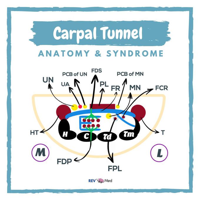
Carpal Tunnel Anatomy & Syndrome. Straight from our YouTube video! @rev.med Link in bio! Anatomy! Follow the diagram in the pic above, or simply go over to YouTube and check out our video! We make it VERY simple. Don’t forget to subscribe! Key: UN: Ulnar Nerve UA: Ulnar Artery PCB of UN: Palmar cutaneous branch of the Ulnar Nerve FDS: Flexor Digitorum Superficialis muscle PL: Palmaris Longus muscle FR: Flexor Retinaculum PCB of MN: Palmar cutaneous branch of the Median Nerve MN: MEDIAN NERVE FCR: Flexor Carpi Radialis muscle FDP: Flexor Digitorum Profundus muscle FPL: Flexor Pollicis Longus muscle T: Thenar muscles HT: Hypothenar muscles BONES: H: Hamate C: Capitate Td: Trapezoid Tm: Trapezium @rev.med on Instagram for more! Clinical! One of the main significance of the Carpal Tunnel drawing is the Carpal Tunnel syndrome which is the compression of the median nerve. One of the ways Carpal Tunnel syndrome can come about is: overuse, thickening of the synovial sheath and tendon, nerve damage, and much more. Clinical manifestation of the Carpal Tunnel syndrome is ‘paresthesia’. In the laymen term it is referred to as limbs falling asleep,and feelings of pins and needles. Know this diagram! This is a fantastic way to learn Anatomy! Subscribe now & make learning EASY!
