


This is an aortic valve with severe aortic stenosis.
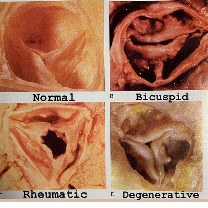
You’ve probably seen my TAVR posts about treating aortic stenosis. Well here is what actual stenotic aortic valve looks like. This was excised from a patient during an open aortic valve replacement. You can see three leaflets, and how it is a thickened abnormal valve with a lot of calcium deposits. This senile calcific degeneration occurs over years and eventually leads to valve narrowing and failure that requires replacement by either open surgical valve replacement or TAVR. Other reasons for aortic stenosis include a congenital valve abnormality such as a bicuspid valve, and rheumatic heart disease. . . 2️⃣ Swipe see this valve specimen with the leaflets and calcium labeled. 3️⃣ Swipe to see a comparison of what a normal valve, bicuspid stenosis, rheumatic stenosis, and calcific degenerative stenosis valve looks like (courtesy of Braunwald)
Source: https://www.instagram.com/p/Bqi_ByOhVBt/?utm_source=ig_share_sheet&igshid=tzfm25ftz98tBreastfeeding and Medications IIEffects of sugar on teethNeurofibromatosisSupraventricular TachycardiaWomen’s Reproductive Health & Daily WellnessGelatine Sculpt | Official WebsiteBurnSlim Official | Support Your Weight Management Journey
