


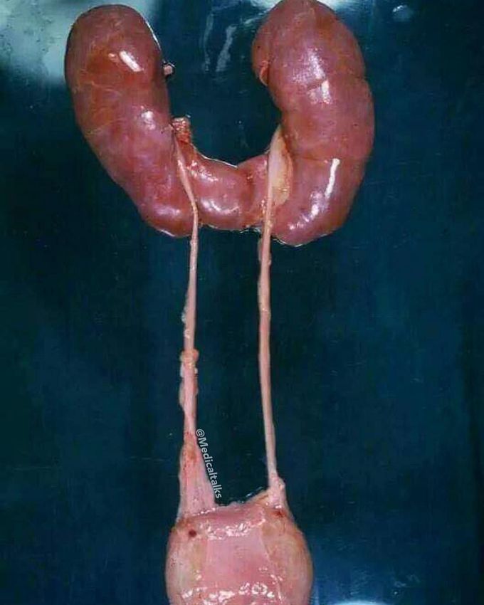
Horseshoe kidney
This is the most common renal anomaly, occurs in one of every 400. Horseshoe kidney is a rental fusion anomaly that consists of two distinct functioning kidneys on each side of the midline, connected at the lower poles by an isthmus (the fused part) of functioning renal parenchyma or fibrous tissue that crosses the midline of the body. Fusion is thought to occur before the kidneys ascend from the pelvis to their normal dorsolumbar position. This is usually between the 5th and 9th weeks of gestation. It often presents together with other congenital anomalies, particularly ureteral and collecting system abnormalities. Horseshoe kidneys are, in themselves, asymptomatic and thus they are usually identified incidentally (eg, routine ultrasound). They are however prone to a number of complications as a result of poor drainage, which may lead to clinical presentation such as pain and/or hematuria due to obstruction or infection. They are at increased risk for infection because of the increased likelihood of urinary stasis. Also susceptibility of trauma and risk of development of renal calculi and transitional cell carcinoma of the renal pelvis. The majority of patients have an excellent prognosis without any therapeutic intervention. It is however important to recognise their presence prior to abdominal surgery or renal intervention for one of their many complications.
Hemodynamic stimuli&nonhemodynamic stimuliEffects of sugar on teeth
