


Zunaira saleh10 months ago
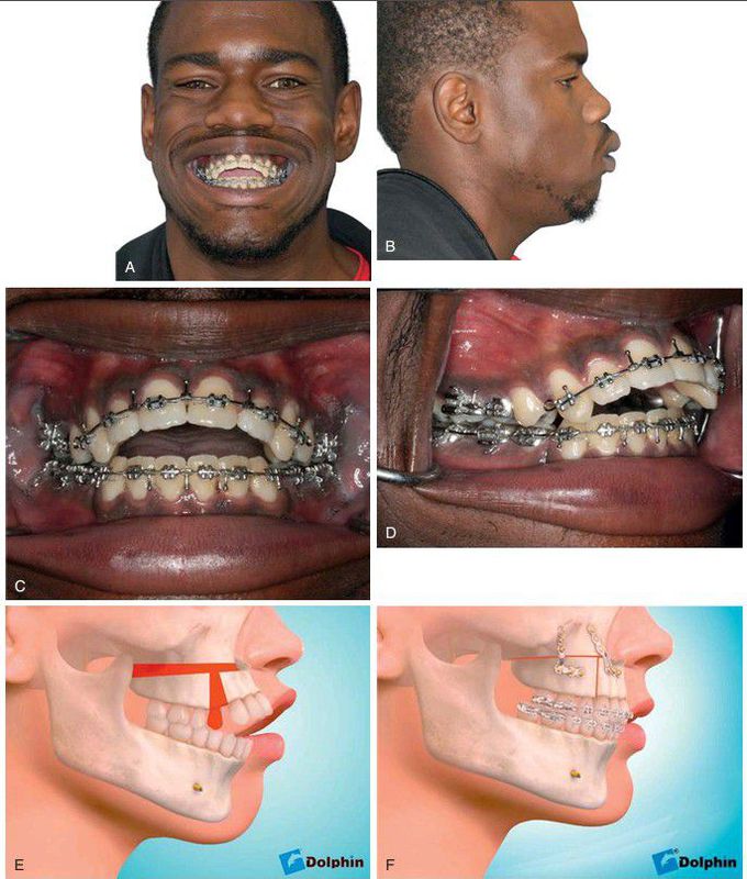
Segmental maxillary osteotomy
. (A–B) Preoperative facial appearance demonstrates extreme protrusion of the anterior maxillary segment and the upper lip, decreased nasolabial angle, and decreased lower face height as a result of maxillary vertical deficiency. (C–D) Preoperative occlusion demonstrates protrusive maxillary incisors and extraction space remaining after removal of maxillary premolar teeth bilaterally. (E–F) Segmental maxillary osteotomy with closure of premolar extraction space, retraction of anterior segment of maxilla, and placement of bone graft in posterior maxillary area.
Other commentsSign in to post comments. You don't have an account? Sign up now!
