


Zunaira saleh9 months ago
3D Imaging and Model for Mandibular Assessment
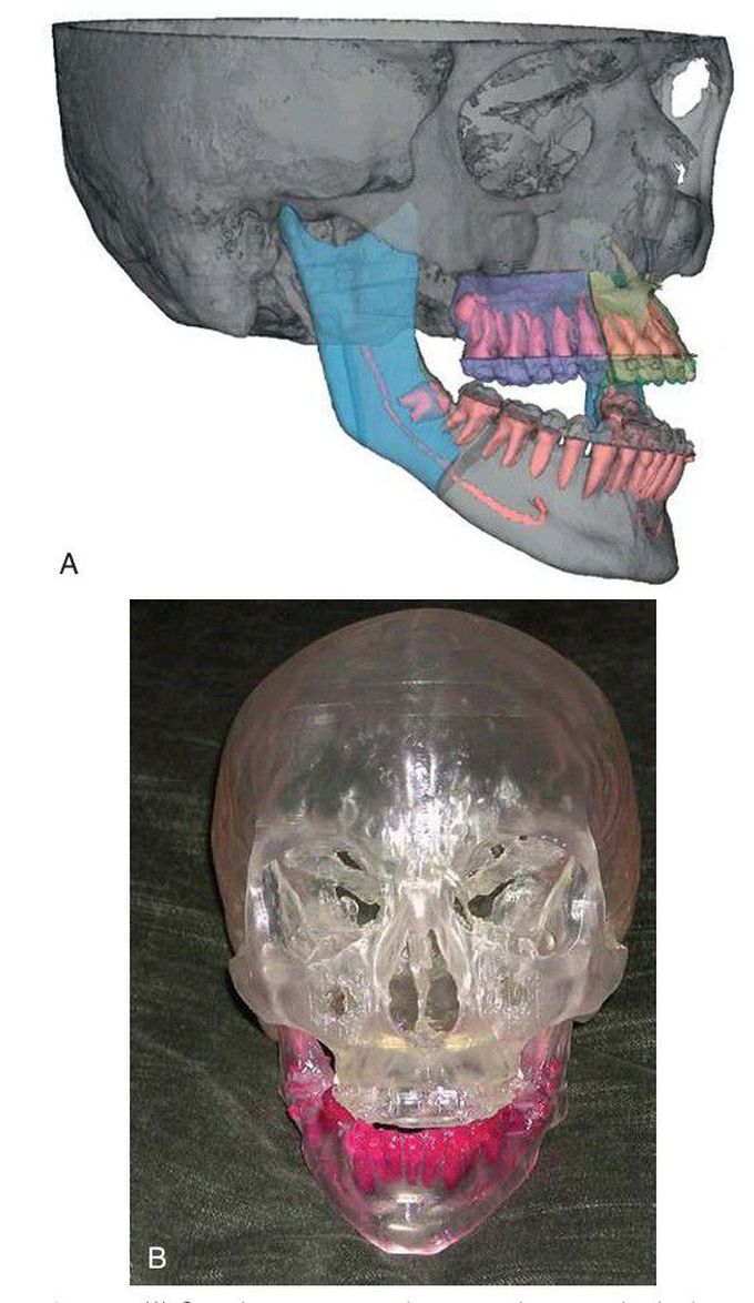
(A) Cone-beam computed tomography scan clearly demonstrating bone deformity in three dimensions with detailed view of skeletal components, tooth root positioning, and location of the inferior alveolar nerve. (B) Stereolithographic model.
Other commentsSign in to post comments. You don't have an account? Sign up now!
