


Zunaira salehabout 1 year ago
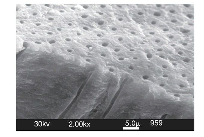
Normal dentin
Scanning electron microscopy (SEM) image of normal dentin showing its unique structure as seen from two directions. At the top is a view of the tubules, each of which is surrounded by peritubular dentin. Tubules lie between the dentin-enamel junction (DEJ) and converge on the pulp chamber. The perpendicular surface at the bottom shows a fracture surface revealing some of the tubules as they form tunnel-like pathways toward the pulp. The tubule lumen normally contains fluid and processes of the odontoblastic cells. (From
Other commentsSign in to post comments. You don't have an account? Sign up now!
