
Zunaira salehabout 1 year ago

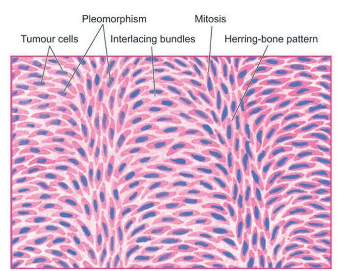
Fibrosarcoma
. Microscopy shows a well-differentiated tumour composed of spindle-shaped cells forming interlacing fascicles producing a typical Herring-bone pattern. A few mitotic figures are also seen.
Other commentsSign in to post comments. You don't have an account? Sign up now!

