
Zunaira salehover 1 year ago

Ewing’s sarcoma.
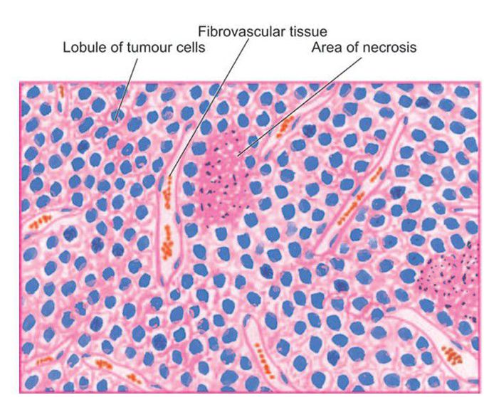
Characteristic microscopic features are irregular lobules of uniform small tumour cells with indistinct cytoplasmic outlines which are separated by fibrous tissue septa having rich vascularity. Areas of necrosis and inflammatory infiltrate are also included. Inbox in the right photomicrograph shows PAS positive tumour cells in perivascular location.
Other commentsSign in to post comments. You don't have an account? Sign up now!

