
Zunaira salehover 1 year ago

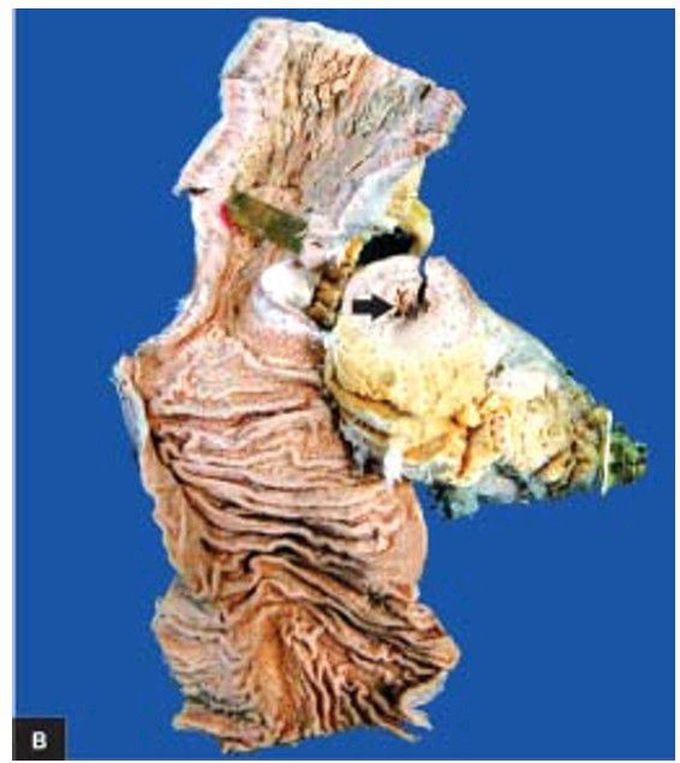
Crohns disease
The specimen of small intestine is shown in longitudinal section along with a segment in cross section. External surface shows increased mesenteric fat, thickened wall and narrow lumen. Luminal surface of longitudinal cut section shows segment of thickened wall with narrow lumen which is better appreciated in cross section (arrow) while intervening areas of the bowel are uninvolved or skipped.
Other commentsSign in to post comments. You don't have an account? Sign up now!

