


Zunaira salehover 1 year ago
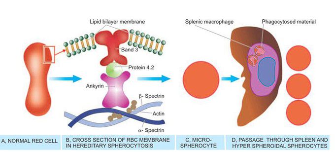
Diagrammatic representation of pathogenesis of hereditary spherocytosis
. A, Normal red cell with biconcave surface and normal size. B, Red cell membrane as seen in cross section in hereditary spherocytosis. Mutations in membrane proteins—α-spectrin, β-spectrin and ankyrin, result in defect in anchoring of lipid bilayer of the membrane to the underlying cytoskeleton. C, This results in spherical contour and small size so as to contain the given volume of haemoglobin in the deformed red cell. D, During passage through the spleen, these rigid spherical cells lose their cell membrane further. This produces a circulating subpopulation of hyperspheroidal spherocytes while splenic macrophages in large numbers phagocytose defective red cells causing splenomegaly.
Other commentsSign in to post comments. You don't have an account? Sign up now!
