
Zunaira salehover 1 year ago

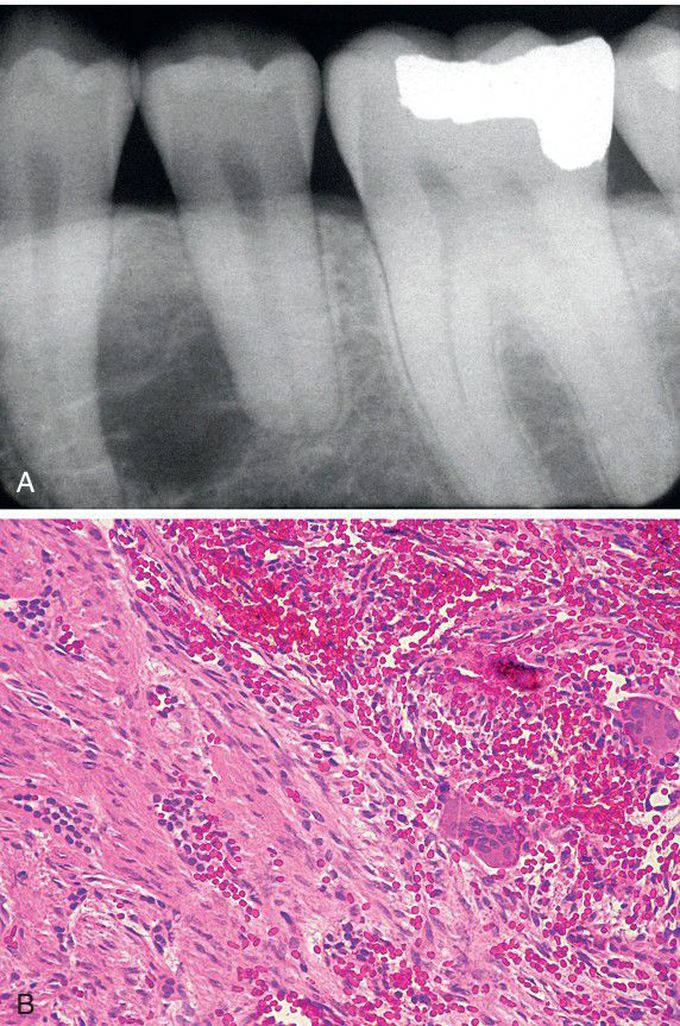
Odontogenic Fibroma (WHO Type) with Associated Giant Cell Granuloma.
A, Unilocular radiolucency between the left mandibular bicuspids. B, Microscopic examination reveals two distinct patterns. On the left, one can see cords of odontogenic epithelium within a fibrous background, consistent with odontogenic f ibroma (WHO type). Typical features of central giant cell granuloma are present on the right side of the field.
Other commentsSign in to post comments. You don't have an account? Sign up now!

