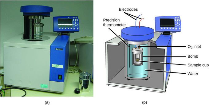


Mostly equipment
[04/10, 11:51 PM] I Love At Times Hacker 💻🔨: COLORIMETRY Colorimetry is a technique which is frequently used in biochemical estimations. It involves the quantitative estimation of color. Thus, a substance to be estimated colorimetrically must be either colored or, most often, capable of forming chromogens through the addition of reagents. The instrument commonly used, known as colorimeter, is in fact absorptiometer. It measures the amount of light absorbed in these estimations. The parts of a colorimeter include a source of light and a device for selecting light of narrow wavelength (Fig. 3.1). The common colorimeters have a set of filters. For example, a green filter absorbs all the component colors of white light except green light which is allowed to pass through. The light that is transmitted through the green filters has a wavelength from 500 to 560 nm. Similarly, other suitable filters can be used to select light of narrow wavelengths. In sophisticated instruments, a diffraction grating or prism is used in place of filters so that the transmitted light has a very narrow bandwidth. The transmitted light passes through a compartment in which the colored solution is placed in a cuvette or test tube. The colored solution absorbs some light and the residual light falls on a detector. The detectors are photosensitive elements which convert the light into electrical signal. The electrical signal generated is directly proportional to the intensity of light falling on the detector. This signal is quantitated by a galvanometer and read as optical density (OD) value. Colored solutions have the property of absorbing certain wavelengths of light and transmitting others. The color of the solution depends upon the transmitted light. For example, a solution of hemoglobin absorbs blue-green light and transmits the complementary red color and hence the solution appears red. In colorimetry, filters are chosen appropriately so that absorption is maximum at the selected wavelength band. Practical Clinical Biochemistry Quantitative Analysis 70 PB Complementary Colors Used in Colorimetry for Colored Solutions Solution color Filter used Peak transmission range (Nm) Bluish Green Red 580 – 680 Blue Yellow 520 – 580 Purple Green 490 – 520 Red Blue-green 470 – 490 Yellow Blue 430 – 470 Yellowish-green Violet 400 – 430 According to Beer’s law, the amount of light transmitted through a colored solution decreases exponentially with increase in concentration of the colored substance. In colorimetric estimation, it is necessary to prepare a ‘reagent blank’, ‘test’ and a ‘standard’. The test solution is made by treating a specified volume of the blood filtrate or other specimen with reagents as mentioned in the procedure. A standard solution is prepared by similarly treating simultaneously, a solution of pure substance whose concentration is known. Since some color comes from the reagents specified in the procedure which is in addition to the color produced by the substance which is desired to be estimated, a blank is also run by similarly treating at the same time a same volume of distilled water. Steps in the Operation of the Photoelectric Colorimeter Place the glass filter recommended in the procedure in the filter slot. Fill the colorimeter tube (cuvette) to about three fourth with the distilled water and place in cuvette slot. Switch ‘on’ the instrument and allow it to warm up for 4 – 5 minutes. Adjust to zero optical density. Take ‘blank’ solution in another tube and with this placed in the cuvette slot, read the optical density (B). Take ‘test’ solution in the cuvette, and as with ‘blank’ read the O.D. (T). Finally take ‘standard’ solution in the cuvette and record O.D. (S). Satisfactory results are obtained only when the O.D. values are in the range 0.1 – 0.7. The reading should be taken with the lighter solution first (lower conc.) followed by the darker solutions (more conc.) in order to avoid erroneous readings due to carryover effect. CALCULATION. Is not available By Vijay Sharma


