


Idiopathic Intracranial Hypertension
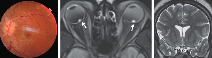
A 25-year-old woman with severe obesity presented to the emergency department with a 1-week history of blurred vision, transient visual obscurations, daily headaches, and intermittent whooshing sounds. Her body-mass index (the weight in kilograms divided by the square of the height in meters) was 57. On neurologic examination, optic-disk swelling and retinal hemorrhages were present in both eyes (Panel A, left eye). The results of visual-field and cranial-nerve testing were normal. Magnetic resonance imaging and venography of the head revealed flattened posterior globes with marked elevation of the optic-nerve heads (Panel B, arrows), an empty sella (Panel C, asterisk), and stenoses of the transverse sinuses without obstruction or thromboses — all of which were suggestive of elevated cerebrospinal fluid pressure. A lumbar puncture was notable for an elevated opening pressure of 55 cm of water (reference range, 10 to 20); the results of the cerebrospinal fluid analysis were normal. A diagnosis of idiopathic intracranial hypertension was made. Idiopathic intracranial hypertension is a disorder associated with obesity that manifests with symptoms resulting from increased intracranial pressure, including headaches, diplopia, visual field defects, and pulsatile tinnitus. Treatment with high-dose acetazolamide was started, and counseling on weight loss was provided. At a 1-month follow-up visit, the patient’s papilledema had decreased, and treatment with acetazolamide had been continued.
