


Abdominal Ectopic Pregnancy
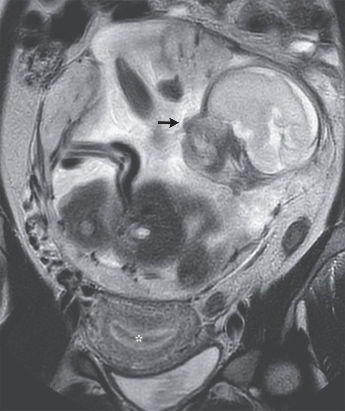
A 37-year-old woman who lived on a remote island presented to the emergency department with a 10-day history of abdominal pain. She had a history of two vaginal deliveries at full term, one miscarriage, and no sexually transmitted infections or previous surgeries. The physical examination was notable for a gravid abdomen. Ultrasonography revealed a thickened endometrium, empty uterus, and abdominal pregnancy at 23 weeks’ gestation. Magnetic resonance imaging of the abdomen subsequently showed a nongravid uterus (asterisk), a normally formed intraabdominal fetus (arrow), and a placenta that attached to the peritoneum above the sacral promontory. A diagnosis of abdominal pregnancy — a rare type of ectopic pregnancy — was made. Owing to the high risk of maternal hemorrhage and fetal demise, the patient was transferred to a tertiary care hospital. At 29 weeks’ gestation, a laparotomy with infant delivery, placental arterial embolization, and partial removal of the placenta was performed. The baby, who had Apgar scores of 2 and 6 at 1 minute and 5 minutes, respectively, was admitted to the neonatal intensive care unit. On postoperative day 12, the remaining placenta was surgically removed. The mother and the baby were discharged home 25 days and 2 months, respectively, after the birth. The mother, who had declined contraception post partum, was lost to follow-up.
