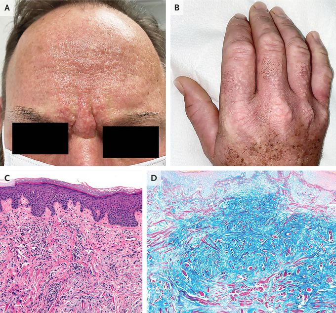
MEDizzy
MEDizzy
Scleromyxedema

Scleromyxedema

A 53-year-old man presented to the rheumatology clinic with a 3-year history of an itchy rash, Raynaud’s phenomenon, dysphagia, and a burning sensation in his hands. Physical examination was notable for firm, greasy papules across his forehead that led to the formation of glabellar grooves (Panel A). There were waxy papules on his hands, with associated skin thickening and finger flexion contractures (Panel B). Similar skin changes were seen on his nose, lips, ears, trunk, and feet. There was no telangiectasia or calcinosis. Sensory neuropathy was present in his hands, arms, and face. Tests of thyroid function were normal. Serum protein electrophoresis with immunofixation identified an IgG-λ monoclonal gammopathy, and a bone marrow biopsy was normal. A subsequent skin-biopsy sample obtained from the right side of the neck showed dermal spindle-cell proliferation, thickened collagen fibers, fibrosis, and perivascular inflammation (Panel C, hematoxylin and eosin stain), as well as increased dermal mucin deposition (Panel D, colloidal iron stain). A diagnosis of scleromyxedema was made. Scleromyxedema is a primary cutaneous mucinosis typically associated with a paraproteinemia. This type of sclerosing skin disorder may cause extracutaneous symptoms, as was seen in this patient. Although infusions of intravenous immune globulin provided minimal relief initially, treatment with lenalidomide resulted in abatement of symptoms and reduction in paraproteinemia after 4 months.

3
Top rated comment
11 months ago

Super sir

Other commentsSign in to post comments. You don't have an account? Sign up now!
Related posts
Long BonesFlat Bones
Stomach
Foot X-ray AnatomyAnatomy of liverAbdominal aortic aneurysmImage of trachea blocked by chewing gum### Anatomy of the Heart: Summary The heart is a muscular organ responsible for pumping blood throughout the body, supplying oxygen and nutrients while removing waste products. It is located in the thoracic cavity, between the lungs, and is roughly the size of a fist. #### 1. **Structure:** - **Chambers:** The heart has four chambers: - **Right Atrium:** Receives deoxygenated blood from the body via the superior and inferior vena cavae. - **Right Ventricle:** Pumps deoxygenated blood to the lungs through the pulmonary arteries. - **Left Atrium:** Receives oxygenated blood from the lungs via the pulmonary veins. - **Left Ventricle:** Pumps oxygenated blood to the rest of the body through the aorta. - **Valves:** The heart contains four main valves that ensure unidirectional blood flow: - **Tricuspid Valve:** Located between the right atrium and right ventricle. - **Pulmonary Valve:** Located between the right ventricle and pulmonary arteries. - **Mitral (Bicuspid) Valve:** Located between the left atrium and left ventricle. - **Aortic Valve:** Located between the left ventricle and the aorta. #### 2. **Wall Layers:** - **Epicardium:** The outer layer, also known as the visceral pericardium. - **Myocardium:** The thick, muscular middle layer responsible for contraction. - **Endocardium:** The inner layer that lines the chambers and valves. #### 3. **Blood Supply:** - The heart receives its blood supply from the coronary arteries, which branch off the aorta. The **right coronary artery** supplies the right side of the heart, while the **left coronary artery** branches into the left anterior descending and circumflex arteries, supplying the left side. #### 4. **Electrical System:** - The heart's rhythmic contractions are controlled by its electrical conduction system, which includes: - **Sinoatrial (SA) Node:** The natural pacemaker located in the right atrium. - **Atrioventricular (AV) Node:** Receives impulses from the SA node and transmits them to the ventricles. - **Bundle of His and Purkinje Fibers:** Distribute the electrical impulse throughout the ventricles, leading to contraction. #### 5. **Function:** - The heart functions in two primary circulatory loops: - **Pulmonary Circulation:** Moves deoxygenated blood from the right side of the heart to the lungs for oxygenation. - **Systemic Circulation:** Distributes oxygenated blood from the left side of the heart to the rest of the body. The anatomy of the heart is crucial for its role in maintaining effective circulation and overall cardiovascular health. Understanding its structure helps in diagnosing and treating various heart conditions.

Recent MCQs


Show more MCQs

Recent flashcard sets


Show more flashcards