


Nailfold Capillaroscopy in Rheumatic Disease
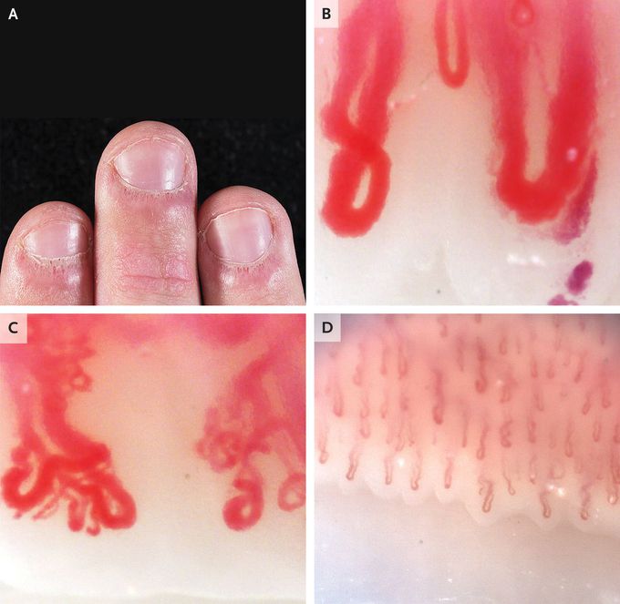
A 19-year-old man presented to the rheumatology clinic with a 3-year history of Raynaud’s phenomenon and a 1-year history of fatigue, rash on the face and hands, and pain in the finger joints. On physical examination, there were erythematous plaques on the metacarpophalangeal and interphalangeal joints, without associated synovitis. The nailbeds had periungual erythema, cuticular hypertrophy (also known as “ragged cuticles”), and prominent dilated capillary loops (Panel A). Erythema was also present on the patient’s hairline, nasolabial folds, and periorbital region. The results of strength testing were normal. Nailfold capillaroscopy was performed. Nailfold capillaroscopy is a bedside assessment of nailbed microcirculation that is performed with the use of a handheld ophthalmoscope, a dermatoscope, a wide-field microscope, or a videocapillaroscopy probe. In this patient, nailfold videocapillaroscopy revealed giant nailfold capillaries with an apical diameter of more than 50 μm (reference value, <15) (Panel B), branched and bushy neovascularized capillary loops (Panel C), and markedly reduced capillary density; for comparison, Panel D shows normal findings on nailfold capillaroscopy. The patient’s nailbed findings, in combination with his symptoms, were suggestive of an underlying rheumatic condition. After serologic evaluation and a skin biopsy, a diagnosis of clinically amyopathic dermatomyositis was made, and the patient’s symptoms abated after immunosuppressive therapy was initiated.
