MEDizzy
MEDizzy
Medication-Related Osteonecrosis of the Jaw

Medication-Related Osteonecrosis of the Jaw

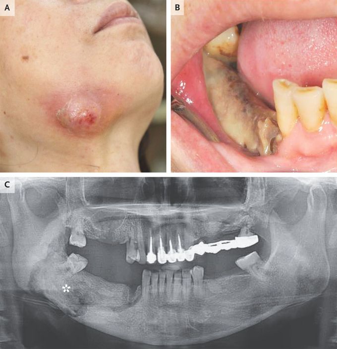
A 54-year-old woman with metastatic breast cancer presented to the oral surgery clinic with a 4-month history of right jaw pain. For the past 10 months, she had been receiving denosumab at a dose of 120 mg every 4 weeks to prevent complications related to bony metastases. Physical examination was notable for swelling over the right jaw from an oral cutaneous fistula that connected to the mandible (Panel A). An area of exposed, necrotic bone was seen inside the mouth in an edentulous region of the right mandible (Panel B). She had several missing teeth and periodontitis. A panoramic radiograph showed an ill-defined radiolucent border with sequestrum over the right mandible (asterisk, Panel C). A diagnosis of medication-related osteonecrosis of the jaw complicated by secondary infection with extraoral fistula formation was made. Medication-related osteonecrosis of the jaw is an uncommon but severe complication of treatment with osteoclast inhibitors, such as denosumab and bisphosphonates. Risk factors include oral surgery, poor oral hygiene, and dental disease. It does not commonly occur with the use of antiresorptive medications at lower doses for conditions such as osteoporosis. The patient underwent surgical débridement and sequestrectomy, during which time treatment with denosumab was discontinued, and antimicrobials and oral rinses were administered. Denosumab therapy was resumed, and at the 1-year follow-up, her symptoms had resolved.

2
Other commentsSign in to post comments. You don't have an account? Sign up now!
Related posts
Flat Bones
Jaw cystsStomach
Foot X-ray AnatomyAnatomy of liverAbdominal aortic aneurysmImage of trachea blocked by chewing gum### Anatomy of the Heart: Summary The heart is a muscular organ responsible for pumping blood throughout the body, supplying oxygen and nutrients while removing waste products. It is located in the thoracic cavity, between the lungs, and is roughly the size of a fist. #### 1. **Structure:** - **Chambers:** The heart has four chambers: - **Right Atrium:** Receives deoxygenated blood from the body via the superior and inferior vena cavae. - **Right Ventricle:** Pumps deoxygenated blood to the lungs through the pulmonary arteries. - **Left Atrium:** Receives oxygenated blood from the lungs via the pulmonary veins. - **Left Ventricle:** Pumps oxygenated blood to the rest of the body through the aorta. - **Valves:** The heart contains four main valves that ensure unidirectional blood flow: - **Tricuspid Valve:** Located between the right atrium and right ventricle. - **Pulmonary Valve:** Located between the right ventricle and pulmonary arteries. - **Mitral (Bicuspid) Valve:** Located between the left atrium and left ventricle. - **Aortic Valve:** Located between the left ventricle and the aorta. #### 2. **Wall Layers:** - **Epicardium:** The outer layer, also known as the visceral pericardium. - **Myocardium:** The thick, muscular middle layer responsible for contraction. - **Endocardium:** The inner layer that lines the chambers and valves. #### 3. **Blood Supply:** - The heart receives its blood supply from the coronary arteries, which branch off the aorta. The **right coronary artery** supplies the right side of the heart, while the **left coronary artery** branches into the left anterior descending and circumflex arteries, supplying the left side. #### 4. **Electrical System:** - The heart's rhythmic contractions are controlled by its electrical conduction system, which includes: - **Sinoatrial (SA) Node:** The natural pacemaker located in the right atrium. - **Atrioventricular (AV) Node:** Receives impulses from the SA node and transmits them to the ventricles. - **Bundle of His and Purkinje Fibers:** Distribute the electrical impulse throughout the ventricles, leading to contraction. #### 5. **Function:** - The heart functions in two primary circulatory loops: - **Pulmonary Circulation:** Moves deoxygenated blood from the right side of the heart to the lungs for oxygenation. - **Systemic Circulation:** Distributes oxygenated blood from the left side of the heart to the rest of the body. The anatomy of the heart is crucial for its role in maintaining effective circulation and overall cardiovascular health. Understanding its structure helps in diagnosing and treating various heart conditions.

Recent MCQs


Show more MCQs

Recent flashcard sets


Show more flashcards