


Gastrointestinal Kaposi’s Sarcoma
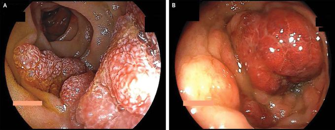
A 54-year-old man with no known medical history presented to the emergency department with a 10-day history of bloody diarrhea and a 3-month history of progressive, red skin lesions. Physical examination revealed cachexia; erythematous papules on the face, torso, and legs; edema of the legs and feet; purplish nodules in the posterior oropharynx; and a distended abdomen. Results of laboratory studies were notable for a positive test for human immunodeficiency virus, a CD4 count of 45 cells per cubic millimeter (reference range, 300 to 1400), and a viral load of 5.5 million copies per millimeter. Computed tomography of the whole body showed a pleural effusion on the right side, pericardial effusion, ascites, diffuse lymphadenopathy, and diffuse thickening of the bowel wall. An upper endoscopy and colonoscopy were performed. Erythematous, polypoid lesions were seen throughout the gastrointestinal tract, from the esophagus to the rectum (Panel A, duodenum; Panel B, sigmoid colon). Biopsy specimens from multiple lesions showed features consistent with Kaposi’s sarcoma. A diagnosis of Kaposi’s sarcoma with cutaneous and gastrointestinal involvement was made. During a prolonged hospital course, the patient also received a diagnosis of cytomegalovirus colitis, and coronavirus disease 2019–related pneumonia. The patient died on hospital day 32.

Living with HIV was one of the hardest experiences of my life. The fatigue, the emotional toll, and the uncertainty about the future weighed on me every single day. I had tried many treatments and medications, but nothing seemed to restore my health or energy the way I hoped.Out of both hope and desperation, I came across NaturePath Herbal Clinic. At first, I was skeptical but something about their natural approach and the powerful stories I read gave me the courage to try one more time.I began their herbal treatment program, and within a few weeks, I noticed small but meaningful changes more energy, better sleep, and a stronger immune system. Over the months, those improvements only grew. Today, I can truly say my life has changed. I feel healthier, more balanced, and finally in control of my well-being again.This isn’t just a testimony it’s a heartfelt recommendation to anyone living with HIV or any chronic condition. Don’t give up hope. I’m so grateful I gave NaturePath Herbal Clinic a chance. Visit their website to learn more: www.naturepathherbalclinic.com Email: info@naturepathherbalclinic.com
