


Iron Deposition from a Retained Intraocular Foreign Body
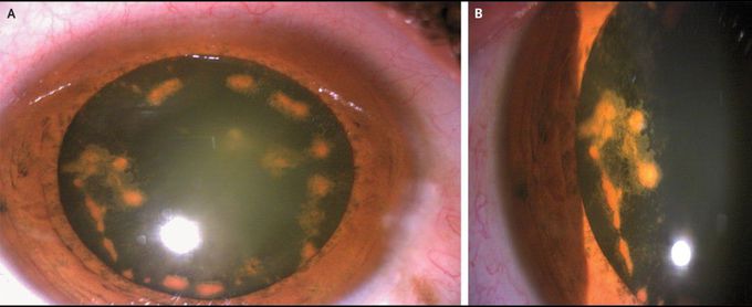
A 55-year-old man presented to the ophthalmology clinic with a 4-month history of decreased vision and floaters in his right eye. Six months before presentation, he was hammering an iron nail without eye protection when something struck his eye. On current examination of the right eye, the best visual acuity was limited to perception of hand motions close to his face. Slit-lamp examination of the right eye showed conjunctival hyperemia, a multicolored iris, and a pigmented cataract with dark-brown anterior subcapsular opacities (Panel A; Panel B shows a magnified view). A small foreign body in the inferonasal peripheral vitreous was also seen. An examination of the left eye was normal. A diagnosis of an intraocular foreign body with associated iron deposition in the eye — also known as ocular siderosis — was made. Ocular siderosis is a pigmentary, degenerative process in which iron deposition in eye structures leads to free-radical damage and subsequent vision loss. The patient underwent a combined phacoemulsification and intraocular lens implant, pars plana vitrectomy, and removal of the foreign body (an iron chip). At a 6-month follow-up visit, visual acuity in the patient’s right eye had improved to 20/40, although electroretinography revealed persistent rod and cone dysfunction.
